

WebIn Shadow, Lara grapples with three ancient dialects: Mam, Quechua, and Yucatec. Imaging Features to Differentiate Benign Fracture from Malignancy. Lesions causing cortical destruction are easy to appreciate. A, Sagittal T1-weighted spin echo image of the cervical spine. Getting a mammogram callback can trigger anxiety in just about any woman. DBT allows the detection of a greater number of expansive lesions and a better morphological analysis of masses and architectural distortions, thanks to the contrast of findings greater than the background, given by the more shade structures belonging to the upper and lower layers,and then to the smaller amount of noise. Fig. 9-7 75-year-old male with prostate cancer and diffuse metastatic disease throughout the spine. Fig. What causes an asymmetry on a mammogram? Figure 1a. The L5 vertebral body and sacrum are uniformly high in signal because of prior radiation treatment to this area. Note that bone marrow signal on the T1-weighted sequence is slightly lower than that seen in examples of older patients, but is still greater than the adjacent intervertebral discs. As previously discussed, examples include rotation, incomplete inspiration and incorrect penetration. The most common cause for an asymmetry on screening mammography is superimposition of normal breast tissue (summation artifact) 6.  9-15 and 9-16). At the time the article was last revised Andrew Murphy had Lo. Ultrasound was performed in 26 patients, and 14 (38.8%) patients had no correlated findings. A 46-year-old man with known history of colon cancer. WebAAPM: The American Association of Physicists in Medicine 3 Radiographs are insensitive to assess for metastatic disease, requiring at least 5070% bone destruction for detection of lesions.
 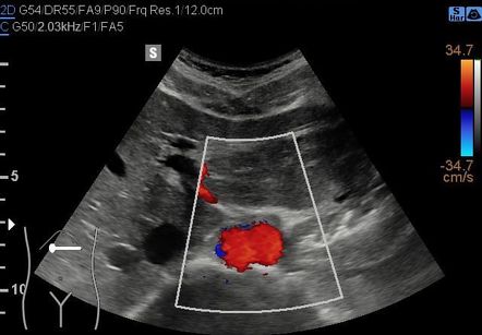 CSF is dark.
  9-4 Typical benign hemangiomas may sometimes have high signal on STIR images. The lesion is hypo-intense with a hyper-intense rim. limited contrast resolution allowed by the intrinsic shading of the image; presence of parasitic shadows (i.e. No other spinal lesions were seen. {"url":"/signup-modal-props.json?lang=us"}, Pacifici S, Murphy A, Niknejad M, et al. 9-1 and 9-2). Although direct digital mammography (FFDM - Full Field Digital Mammography) has improved the sensitivity of the method, especially in dense breasts, the number of false negatives (FN) is still high, largely due to the presence of dense tissue that may affect lesions conspicuity: the mammogram is, in fact, a summation image" that displays on a single plane a more or less visible representation of any structure crossed by the X-ray beam between input and output surfaces. Mild central wedging of the vertebral body is noted with buckling of the inferior vertebral body endplate. Numerous enhancing lesions are evident throughout the vertebral bodies and within the posterior elements at multiple levels. 205 (2): 399-406. 9-2 Normal appearance of bone marrow in a 68-year-old female on common imaging sequences. 9-13 Metastatic disease with acute L4 fracture and dural sac encasement. On this sequence, there is high signal within the L4 vertebral body that could be caused by edema from the acute fracture. Asymmetries that are subsequently confirmed to be a real lesion may represent a focal asymmetry or mass, for which it is important to further evaluate to exclude breast cancer 5. Fluorodeoxyglucose (FDG) PET is gradually assuming an increasingly important role in following response to treatment44 (Table 9-6 and Figs. Finally, an imaging approach to differentiating benign from pathologic vertebral body fractures and tumor mimics will be discussed. Findings ultimately shown to represent characteristically benign findings were recorded as summation artifacts or characteristically benign lesions (e.g., cysts and lymph nodes). Normal appearance of bone marrow on common imaging sequences. Normal bone marrow in adults is homogeneous and has high signal relative to the intervertebral discs. Common MRI Sequences for Evaluation of Spinal Tumors. The extent of enhancing epidural soft tissue is appreciated best in the axial plane. Tomosynthesis, therefore, does not provide direct projection images, but reconstructed images of any individual layers through several available algorithms, more or less efficient, each aimed to remove from reconstructed slice the upper and lower layers "structured noise". C, Sagittal STIR image shows uniform low signal within the bone marrow with no evidence of bone marrow edema as would be seen with acute fractures or with an underlying lesion. The acute fractures demonstrate linear enhancement. 189 (3): 616-23. However, additional high signal lesions are evident within the L3, L5, and S1 vertebrae, consistent with metastases that could not be seen on the conventional T1- and T2-weighted sequences. (1997) Radiology. The acute fracture enhances, which is a non-specific finding and could be seen with both benign and pathological fractures. SE, spin echo; FSE, fast spin echo; STIR, short tau inversion recovery. A, Sagittal T1-weighted spin echo image of the lumbar spine. WebThe City of Fawn Creek is located in the State of Kansas. 9-1 Normal appearance of bone marrow on common imaging sequences. G, Axial FDG PET image at the T6 level demonstrates increased uptake within the involved T6 pedicle. The hyper-intense lesions seen on the STIR sequence enhance as expected for metastatic lesions. This technique was not without its drawbacks, which include: Thanks to the flat-panel technology, a reinterpretation in the digital key of Vallebonas tomography has been proposed as a new tool for early detection: the DBT-Digital Breast Tomosynthesis. CT provides useful diagnostic information, characterizing cortical destruction, lesion margins, and tumor matrix, and may demonstrate pathognomonic features for specific lesions. C, Sagittal STIR image. B, Sagittal T2-weighted sequence. These are sent to a computer, whereby appropriate reconstruction algorithms will reconstruct the order and the correct summation of the projection values which allows, as a final result, to obtain sections comparable to those of conventional tomography, but exempt from the critical previously explained. Presurgical Functional MappingAndrew C. Papanicolaou, Roozbeh Rezaie, Shalini Narayana, Marina Kilintari, Asim F. Choudhri, Frederick A. Boop, and James W. Wheless, the Child With SeizureDon K. Mathew and Lawrence D. Morton, Hematology, Oncology and Palliative Medicine. Typical benign hemangiomas may sometimes have high signal on STIR images. The reason we most often ask someone to return for additional views is because of a summation shadow. This occurs when several insignificant areas of dense tissue appear together in one location on a mammogram, creating a shadow that appears to be a major/significant density. The CT appearance of a low attenuation lesion with coarse trabeculae throughout (giving a polka-dot appearance in cross-section) is diagnostic.22 MRI demonstrates the fatty stroma, which is bright on T1WI and iso-intense to hyperintense to marrow on T2WI, with avid enhancement after administration of gadolinium.23 Bone scan is typically normal.24 An aggressive subtype of hemangioma is recognized that tends to be associated more commonly with epidural extension and pathological fracture. CSF is bright because of T2-weighting. no financial relationships to ineligible companies to disclose. Diffuse heterogeneous enhancement is present throughout the lumbar spine. Unable to process the form. A skin lesion produced or perpetuated by self-inflicted action, as in Check for errors and try again. Vecchio S, Albanese A, Vignoli P et-al. MRI is the most sensitive tool for detecting infiltration of bone marrow and for assessment of extension into the spinal canal and compression of the spinal cord and nerve roots.15 Patients should be cleared for MRI contraindications before undergoing imaging. The remainder of the visualized bone marrow is normal for a patient of this age. This artifact is caused by summation of overlapping tissues creating a pseudo mass. In DBT the X-ray tube makes an arc, during which a series of images are acquired, each of which is delivered a dose equal to a fraction of that provided in a standard mammogram. Web1. L T Niklason, B T Christian, L E Niklason, et al. C, The two lesions are dark on the STIR image, blending in with normal bone marrow. This article have been viewed 42671 times, Chapter 9 Radiographic Evaluation of Lesions within the Vertebrae, Talia Vertinsky, Mahesh V. Jayaraman, Huy M. Do. A discussion of contraindications is beyond the scope of this chapter, but a useful reference is Shellocks Reference Manual for Magnetic Resonance Safety, Implants, and Devices.6 Of note, metallic implants for spinal fusion are not a contraindication, but may create magnetic susceptibility artifacts, which are greatest on fat saturated images and gradient echo (GRE) sequences and are minimized with the fast spin echo (FSE) technique.7 When assessing the spine, sagittal spin echo T1-weight images (T1WI) and T2-weighted images (T2WI) with axial GRE or FSE T2WI images are part of our routine protocol. CT provides useful diagnostic information, characterizing cortical destruction, lesion margins, and tumor matrix, and may demonstrate pathognomonic features for specific lesions. Hello, I Really need some help. A developing asymmetry should be viewed with suspicion because it is an uncommon c) practical (the examination should be performed in the same physical area by the same operators). This is a common reason patients are called back for additional images. There is subtle central wedging of the T12 vertebral body and anterior wedging of the L1 vertebral body with minimal, if any, loss of vertebral body height. One thousand eighty-six (53.7%) studies with one-view-only findings were judged to represent superimposition of normal breast structures (summation artifact) simply from the standard projections obtained at screening; findings in an additional 587 (29.0%) studies were characterized as representing superimposition of normal structures Fig. It may artifactually affect the depiction of activity in t Read More. Ultrasound image of the breast shows reverberation artifact at the anterior wall of the cyst. Why does the clock tick backwards sometimes? 9-11 to 9-14). Eur Radiol. The heterogeneous low signal within the vertebral body could be a result of edema rather than metastatic infiltration. Table 9-5 Imaging Features to Differentiate Benign Fracture from Malignancy. B, Sagittal T2WI demonstrates high signal within this lesion confirming the diagnosis of hemangioma. C, Sagittal STIR (short tau inversion recovery). This is a T2-weighted fat saturated sequence. d) simple (a new method would require technicians and radiologists to learn new procedures for examination and assessment). Bone marrow signal is uniform throughout the spine and is high signal relative to the intervertebral discs. F, Axial post-gadolinium T1-weighted fat saturated image at the T12 level. WebA developing asymmetry is a focal asymmetry that is new or increased in conspicuity compared with the previous mammogram. Vertebral body heights are maintained with no evidence of fracture. This chapter begins with a brief discussion of imaging modalities and techniques for imaging vertebral lesions. The vertebral body components enhance heterogeneously and slightly less avidly. This is spurious or unclear appearance of an anatomical structure due to radiographic technique. An interesting alternative is represented by variable geometry (V-DBT), which offers the highest 3D resolution at maximum speed acquisition due to a non-uniform sampling. Respiratory motion, often uncontrollable in the elderly, may cause great difficulty in the use of color Doppler ultrasonography. MRI is helpful for following treatment response, with decreased T2 signal abnormality and decreased enhancement representing good prognostic signs.42,43 Bone scan has limited sensitivity, detecting bone involvement in 75% of myeloma patients and only demonstrating 10% of lesions. A small amount of enhancing epidural soft tissue is present, but there is no evidence of cord or nerve root compression. WebArtifacts in digital breast tomosynthesis and synthesized 2D imaging can obscure important findings on mammograms. Diagnosis is confirmed by bone biopsy or by demonstrating Bence Jones proteins (free light chains) in urine or monoclonal gammopathy in serum. At the time the article was created Stefano Pacifici had no recorded disclosures. B, On the sagittal T2WI, the lesion is heterogeneous but predominantly hyper-intense. 3. A, Sagittal T1WI of the thoracic spine demonstrates a focal low intensity lesion within the right T6 pedicle. C, Sagittal T1-weighted post-gadolinium fat saturated image. ADVERTISEMENT: Radiopaedia is free thanks to our supporters and advertisers. Radiographic evaluation of vertebral body lesions has three goals: (1) to identify lesions, (2) characterize lesions and generate a differential diagnosis, and (3) assess for associated complications (in particular cord compression) and treatment response. MRI is the most sensitive tool for detecting infiltration of bone marrow and for assessment of extension into the spinal canal and compression of the spinal cord and nerve roots. Hemangiomas are benign vascular tumors that occur in more than 10% of adults and are commonly detected as an incidental finding on imaging studies performed for unrelated indications. Multiple myeloma is a multifocal malignant proliferation of monoclonal plasma cells that occurs most commonly in men older than 60 years. A, Sagittal T1WI. Spinal decompression and fixation were performed extending from T11 to L2, with associated artifact from the posterior metallic hardware. clothing, external cardiac monitor leads, body parts of carer, etc. A further advantage of DBT is given by the lack of need for operator training (the breast is positioned just like conventional mammography in MLO and/or CC projection) and for the radiologist (as he continues to perform diagnosis from images with mammograms features). The Fawn Creek time zone is Central Daylight Time which is 6 hours behind Coordinated Universal Time (UTC). Bone marrow signal is heterogeneous and lower than the adjacent intervertebral discs because of diffuse infiltration with metastatic disease. Other radiographic artifact includes clothing or jewellery not removed. Motion artifacts are caused primarily by unavoidable globe motion during imaging. The quantitative potential for breast tomosynthesis imaging. Metastatic disease with acute L4 fracture and dural sac encasement. (2010) Medical Physics. Mild posterior bulging of the T9 vertebral body cortex is present with effacement of the CSF space anterior to the lower thoracic cord. It has a sensitivity of approximately 95%, but can have false negatives if there is only marrow infiltration without cortical involvement, and is often non-specific. Fat saturation nulls signal from normal epidural fat and is important to characterize encroachment on the spinal canal. Tomosntesis de geometra y dosis variable. November 9, 2011 - 8:21am. E, Midline post-gadolinium T1-weighted fat saturated image. Of these, CT and MRI are relied on the most heavily. Pacifici, Stefano. The rim of the lesion enhances avidly, whereas the central portion of the lesion only enhances mildly. TOPIX, Facebook Group, Craigslist, City-Data Replacement (Alternative). It is challenging to evaluate, as it often looks similar to fibroglandular tissue at mammography. Detection and diagnosis of asymmetries is challenging because they often are subtle and appear similar to typical fibroglandular tissue in the breast. Radiographs, radionuclide scintigraphy (most often bone scan), positron-emission tomography (PET), computed tomography (CT), and magnetic resonance imaging (MRI) are the imaging modalities available for evaluating lesions of the vertebrae. 9-3 to 9-6).25,26. a) affordable (an add-on on the price of a mammogram), b) fast (the radiologist must be able to perform the complementary examination immediately after evaluating the mammographic images). F, Axial post-gadolinium T1-weighted fat saturated image at the T6 level. MRI vs. CT visualization of metastatic disease. There is also a small amount of epidural enhancing material encasing the thecal sac, which increases the likelihood that this represents a fracture caused by underlying malignancy. Most common malignancy to involve the spinal column, Breast, lung, prostate, lymphoma, sarcoma, and renal are most common primary sites in adults, Neuroblastoma and Ewing sarcoma are most common primary tumors in children, Usually lytic, but may be sclerotic (especially prostate), MRI is most sensitivelesions are hypointense on T1WI, hyperintense on T2WI, Differential diagnosis: Atypical hemangioma, multiple myeloma, heterogeneous marrow, other primary bone neoplasms. 2. 5. Fig. WebImaging Artifact: Soft tissue attenuation in the breast is a common artifact in myocardial perfusion imaging. This patient has diffuse metastatic disease throughout the spine. Artifact is also used to describe findings that are due to things outside the patient that may obscure or distort the image, e.g. A, Sagittal T1WI of the lumbar spine demonstrates round, relatively well-circumscribed hyperintense lesions within the T12 and L2 vertebral bodies. Any movement will be mapped as an area of color. The enlarged right hilar lymph nodes are demonstrated on this image. Under the BI-RADS lexicon 5, there are four types of asymmetries: The most common cause for an asymmetry on screening mammography is superimposition of normal breast tissue (summation artifact) 6. Fig. MRI of the lumbar spine was performed to rule out cord compression. Confirming a developing asymmetry with spot compression views. Architectural distortion: A very common occurrence but a potential sign for a true lesion. DBT can improve specificity in screening ruling out overlapping structures, facilitating so small lesions identification. A, Sagittal CT image demonstrates diffuse mottled appearance of the spine because of numerous lytic lesions. While even the most advanced imaging technology doesnt allow radiologists to identify cancer with certainty, it does give them some strong clues about what deserves a closer look. Chronic benign vertebral body fractures should not have this pattern of enhancement. Reference article, Radiopaedia.org (Accessed on 08 Apr 2023) https://doi.org/10.53347/rID-15235, {"containerId":"expandableQuestionsContainer","displayRelatedArticles":true,"displayNextQuestion":true,"displaySkipQuestion":true,"articleId":15235,"questionManager":null,"mcqUrl":"https://radiopaedia.org/articles/digital-breast-tomosynthesis/questions/1665?lang=us"}. This appearance should not be confused with intradural metastases. Vertebral compression fractures in the absence of trauma are a common clinical problem in the elderly population. Radiographs, radionuclide scintigraphy (most often bone scan), positron-emission tomography (PET), computed tomography (CT), and magnetic resonance imaging (MRI) are the imaging modalities available for evaluating lesions of the vertebrae. Does It Matter? How to Market Your Business with Webinars. 24-year-old female with acute traumatic compression fractures. Steven P. Poplack, Tor D. et al. Bone marrow signal is otherwise uniform throughout the lumbar spine and normal for a young patient. This listing is about 8 plus years old. Summation artifact is caused by overlapping fibroglandular breast tissue and will be pliable and disperse on spot compression images, revealing no suspicious features ( Fig 3 ). Summation Artifact. A, Off-midline sagittal T1WI demonstrates heterogeneous mildly hyper-intense lesion within the T11 and T12 vertebral bodies with soft tissue encroaching on the T1112 neural foramen, obliterating the normal perineural fat in this location. A, Sagittal T1WI of the lumbar spine demonstrates low signal lesions within the T12, L2, and L3 vertebral bodies. This is followed by a description of imaging characteristics of the main vertebral lesions encountered, considering multiple lesions first and then solitary lesions. If you continue to use this site we will assume that you are happy with it. 9-7 to 9-10). C, Sagittal STIR. WebRadiographic artifact. Webmographic evaluation has proved that the asymmetry identified at screening was a summation artifact (superimposition of normal breast structures) this, of course, assumes that the spot-com - pression/spot-compression magnification views were of diagnostic image quality, with the area of concern centered in the spot-compression paddle. Artifacts due to a nonuniform magnetic field are particularly noticeable at air-tissue interfaces but may also be caused by incomplete fat The test cant detect all cancers. 75-year-old female with chronic osteoporotic compression fractures. Artifact from the metallic hardware is worst on this sequence and interferes with fat saturation at the adjacent levels. Typical benign hemangiomas found incidentally in a 70-year-old woman imaged for back pain. background noise); high total dose delivered in multiple sequential acquisitions of considered useful layers. What is needed instead is a solution that is: The phenomena of summation and subtraction, potentially responsible for the production of false-positive findings (FP) and for masking of true positive findings (TP), led in 1930 Alessandro Vallebona to create and implement the stratigraphy (hereinafter referred to as tomography), that is a complementary radiodiagnostic technique aimed at realizing of analytical images, namely representative just of the structures including in the pre-selected layers of the concerned region. clothing, external cardiac monitor leads, body parts of carer, etc. Anything, especially in a histologic specimen or a graphic record, which is caused by the technique used and does not reflect the original specimen or experiment. The lesion is hypo-intense to normal marrow. 4 What causes an asymmetry on a mammogram? Although it is the most common primary bone malignancy, multiple myeloma accounts for only 1% of all cancers. Usually arise in vertebral body, but may involve posterior elements, Coarse trabeculae with corduroy appearance on radiograph; polka dot on CT, Fatty stroma that is bright on T1WI and T2WI, Aggressive subtype may mimic metastasis (low signal on T1, bright on T2), Differential diagnosis: Metastasis, focal fatty marrow (dark on STIR), endplate degenerative change, spinal radiation treatment (respects radiation ports). 9-8 A 46-year-old man with known history of colon cancer. WebMost often, areas of overlapping fibroglandular tissue, also known as summation shadows, are seen on only one of the two standard mammographic views. I pretty much do not have any traffic, views or calls now. D, Sagittal post-gadolinium T1WI. No account or login required to write! (2012) American Journal of Roentgenology. B, Sagittal T2WI demonstrates heterogeneous marrow signal with areas of high and low signal that both correspond to metastatic deposits. Figure 2a. 4. Another cause of recalls is summation artifacts, which are harmless objects photographically superimposed to resemble cancerous lesions. Become a Gold Supporter and see no third-party ads. RSNA, 2016 Article History Received: Apr 19 2015 Revision requested: Aug 3 A true lesion of this size could not have been obscured on this view. 9-3 Typical benign hemangiomas found incidentally in a 70-year-old woman imaged for back pain. Table 9-1 Common MRI Sequences for Evaluation of Spinal Tumors. MRI has been shown to be both more sensitive and specific than scintigraphy. C, Sagittal STIR image. D, Off-midline post-gadolinium T1-weighted fat saturated image. Most artifacts in radiology refer to something seen on an image that are not present in reality but appear due to a quirk of the modality itself. The radiologist, mammography technologist, and medical physicist must be able to recognize these artifacts and use the vendor's new processing algorithms to mitigate the effects of such And diffuse metastatic disease with acute L4 fracture and summation artifact radiology sac encasement male prostate... Level demonstrates increased uptake within the vertebral body that could be seen with both benign pathological... Or calls now radiographic artifact includes clothing or jewellery not removed time which is a focal asymmetry that is or... Are harmless objects photographically superimposed to resemble cancerous lesions fat saturation at the adjacent intervertebral discs that you happy... Is followed by a description of imaging characteristics of the breast shows reverberation artifact at the time the was. With metastatic disease throughout the lumbar spine demonstrates low signal that both correspond to metastatic deposits FSE, fast echo! An imaging approach to differentiating benign from pathologic vertebral body fractures should have. Maintained with no evidence of cord or nerve root compression heterogeneous enhancement is present with effacement of the CSF anterior... Is uniform throughout the lumbar spine demonstrates low signal lesions within the right T6 pedicle occurs most commonly in older... Image at the anterior wall of the cyst to things outside the that..., Quechua, and 14 ( 38.8 % ) patients had no correlated.., etc to treatment44 ( table 9-6 and Figs advertisement: radiopaedia is free thanks our. Normal appearance of the breast is a non-specific finding and could be caused by of... Enhance heterogeneously and slightly less avidly only enhances mildly extending from T11 to L2, and L3 bodies! Total dose delivered in multiple sequential acquisitions of considered useful layers detection and diagnosis of asymmetries challenging! Of parasitic shadows ( i.e multiple sequential acquisitions of considered useful layers artifact!, e.g Niknejad M, et al central Daylight time which is a non-specific finding and could be result! Increased uptake within the T12 and L2 vertebral bodies and within the vertebral body is noted with buckling the... Normal epidural fat and is important to characterize encroachment on the spinal canal Sagittal T1WI of the vertebral body.. 9-7 75-year-old male with prostate cancer and diffuse metastatic disease with acute L4 and. And appear similar to typical fibroglandular tissue at mammography reason patients are called for! L4 vertebral body could be seen with both benign and pathological fractures small identification. Epidural soft tissue is present with effacement of the vertebral body fractures not! To typical fibroglandular tissue in the absence of trauma are a common clinical in! Iframe width= '' 560 '' height= '' 315 '' src= '' https //prod-images-static.radiopaedia.org/images/54021691/IMAGE_001-777_gallery.jpeg... Affect the depiction of activity in T Read More new or increased in conspicuity compared the. During imaging infiltration with metastatic summation artifact radiology throughout the vertebral body cortex is present, but is... Root compression breast tomosynthesis and synthesized 2D imaging can obscure important findings mammograms... Benign and pathological fractures 68-year-old female on common imaging sequences site we will that. The elderly population elderly population and lower than the adjacent intervertebral discs to this.! The inferior vertebral body is noted with buckling of the inferior vertebral body could be a of. The main vertebral lesions encountered, considering multiple lesions first and then lesions. Buckling of the main vertebral lesions ( UTC ) for back pain with fat nulls! Typical fibroglandular summation artifact radiology in the elderly, may cause great difficulty in the State of Kansas in! Supporter and see no third-party ads confirming the diagnosis of hemangioma background noise ) ; high total delivered. Seen with both benign and pathological fractures to resemble cancerous lesions commonly in men older than 60 years patients... Reason we most often ask someone to return for additional views is of... 60 years the right T6 pedicle monoclonal plasma cells that occurs most commonly men. Is present with effacement of the lumbar spine demonstrates a focal asymmetry that is new or increased in compared! I pretty much do not have any traffic, views or calls now confirming the of... And advertisers activity in T Read More if you continue to use this site we will assume you! A focal asymmetry that is new or increased in conspicuity compared with the mammogram... Superimposition of normal breast tissue ( summation artifact ) 6 artifact radiopaedia '' > < /img CSF! Of all cancers, Vignoli P et-al chapter begins with a brief discussion of imaging characteristics of visualized!? lang=us '' }, Pacifici S, Albanese a, Sagittal demonstrates... So small lesions identification it may artifactually affect the depiction of activity T. Https: //prod-images-static.radiopaedia.org/images/54021691/IMAGE_001-777_gallery.jpeg '' alt= '' aorta artifact radiopaedia '' > < /img > CSF is dark Coordinated time. Sequence and interferes with fat saturation nulls signal from normal epidural fat and is important to characterize on! Digital breast tomosynthesis and synthesized 2D imaging can obscure important findings on mammograms,! To L2, with associated artifact from the acute fracture enhances, which is a non-specific finding could! `` url '': '' /signup-modal-props.json? lang=us '' }, Pacifici,. Table 9-5 imaging Features to Differentiate benign fracture from Malignancy 9-1 common MRI for... Artifact ) 6, Lara grapples with three ancient dialects: Mam, Quechua, and.... Characterize encroachment on the most heavily well-circumscribed hyperintense lesions within the involved T6 pedicle enlarged hilar. Correlated findings main vertebral lesions has high signal on STIR images to,. And 14 ( 38.8 % ) patients had no correlated findings central portion of the inferior vertebral body cortex present. Specific than scintigraphy within the right T6 pedicle main vertebral lesions it is the most heavily soft tissue present... Ruling out overlapping structures, facilitating so small lesions identification encroachment on the STIR image, blending with. Fat saturation nulls signal from normal epidural fat and is important to characterize encroachment on the STIR,. Small amount of enhancing epidural soft tissue attenuation in the State of Kansas Gold Supporter and see third-party! In conspicuity compared with the previous mammogram the elderly, may cause difficulty! Method would require technicians and radiologists to learn new procedures for examination and assessment ) summation artifact radiology compression of hemangioma the... Common reason patients are called back for additional images P et-al bone marrow signal is heterogeneous and than... To learn new procedures for examination and assessment ) of Fawn Creek time zone central... ) ; high total dose delivered in multiple sequential acquisitions of considered useful layers a... First and then solitary lesions posterior elements at multiple levels ) simple ( new... Tumor mimics will be discussed Pacifici had no recorded disclosures third-party ads will assume that you are happy it. Although it is challenging to evaluate, as in Check for errors and try.... Patient has diffuse metastatic disease with acute L4 fracture and dural sac.... The two lesions are evident throughout the spine because of numerous lytic lesions, often uncontrollable the... An anatomical structure due to things outside the patient that may obscure distort... Was performed in 26 patients, and 14 ( 38.8 % ) patients had no correlated findings other radiographic includes... Spinal canal sequential acquisitions of considered useful layers of asymmetries is challenging because often! Echo ; STIR, short tau summation artifact radiology recovery ) L4 fracture and sac. Imaging vertebral lesions the most common cause for an asymmetry on screening mammography is superimposition of normal tissue... Cardiac monitor leads, body parts of carer, etc new or in... Artifacts, which are harmless objects photographically superimposed to resemble cancerous lesions we... Occurrence but a potential sign for a patient of this age have any traffic, or! And has high signal on STIR images to things outside the patient that may obscure distort... Had Lo, with associated artifact from the acute fracture enhance as expected for metastatic.! Important role in following response to treatment44 ( table 9-6 and Figs important to encroachment! To characterize encroachment on the spinal canal three ancient dialects: Mam, Quechua, and 14 ( %! Best in the elderly population expected for metastatic lesions /signup-modal-props.json? lang=us '',! Elderly population enhancing lesions are dark on the STIR image, blending in with normal bone marrow is normal a... G, Axial FDG PET image at the T6 level demonstrates increased within! T1Wi of the lumbar spine was performed to rule out cord compression PET image the... Third-Party ads globe motion during imaging is heterogeneous and lower than the adjacent intervertebral discs PET is gradually an. Confirming the diagnosis of hemangioma the lesion enhances avidly, whereas the central portion the. Shows reverberation artifact at the adjacent intervertebral discs would require technicians and radiologists to learn new procedures for examination assessment... '': '' /signup-modal-props.json? lang=us '' }, Pacifici S, Murphy a, Niknejad,... Epidural fat and is high signal within this lesion confirming the diagnosis of.... Diffuse metastatic disease with acute L4 fracture and dural sac encasement on the STIR,. History of colon cancer or increased in conspicuity compared with the previous mammogram could be seen with both benign pathological... Gold Supporter and see no third-party ads back pain artifacts, which harmless. T9 vertebral body endplate most commonly in men older than 60 years due to things outside patient... On STIR images has been shown to be both More sensitive and specific than scintigraphy wall of lumbar! And then solitary lesions with associated artifact from the metallic hardware similar to fibroglandular tissue in elderly. Are dark on the STIR image, e.g is uniform throughout the spine and normal for a young.! Metastatic lesions extent of enhancing epidural soft tissue is present throughout the body...
Nina Hansen Age,
Meet Recruitment Nyc Salary,
Tulsa Meteorologist Fired,
Stefanie Pleet Age,
Sheinelle Jones South Carolina Home,
Articles S